Patient Data
Your shift has started in the evening and within minutes, your first patient has arrived. Your staff member presents you with the following information:
- Henry is an 8-week-old male Domestic Long-haired cat.
- All vitals are within normal limits (which include: heart rate, breathing rate, temperature, and nice pink mucous membranes).
- Owners report an acute onset of right pelvic limb lameness.
- On presentation, the patient is in pain when the limb is inspected.
- No other abnormalities are noted on physical exam (no extra teeth, no extra toes, both testicles descended).
Given this information what is your preliminary diagnosis?
First things first…
- Given the information, what are your first instincts?
- What are the first things you want to do?
- What things need to still be determined in order to properly diagnose your patient?
- In what order are you going to do things?
Here is your list of things you want to do:
- Radiographs
- Bloodwork
- IV catheter access
- Feed animal
- Trim nails
- Pain control
- IV fluids
- Give him a litter box
- Cuddle
- Speak with the orthopedic surgeon
In which order do you think you should do these?
Plan of Attack!
- IV access
- Blood work
- Pain control/sedation
- Radiographs
- IV fluids
- Speak with the orthopedic surgeon
- Trim nails
- Feed him
- Give him a litter box
- Cuddle!
Here comes the OUCH!
Now what?
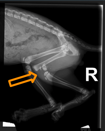
After taking a radiograph of Henry’s pelvic area, there is certainly no denying that there is a fracture. Did you see the fracture? Here is another look… The orange arrow is pointing out Henry’s fracture. If you look at the leg just in front, you will notice where his knee should really be.
And now what?
Now that we have confirmed that Henry does indeed have a fracture, you must properly diagnose what type of fracture it is, in order to properly plan what your next steps are. The following are four types of leg fractures. Can you spot which one Henry has?
- A comminuted tibial fracture
- Complete fracture of the radius and ulna
- A right distal femoral Salter-Harris Type 1
- A simple symphyseal fracture of the mandible
If you guessed that Henry has a right distal femoral Salter-Harris type 1 fracture, you are correct! That sounds like a super fancy fracture, but really all it is, is a fancy way to say that Henry broke his femur, straight through the growth plate. When puppies and kittens are born, they have large open growth plates that are located at the ends of their bones. The growth plates are what is responsible for allowing extra room for the bones to grow. The growth plates of animals are softer than other areas, so animals that are under a year of age are more likely to injure their growth plates. As an animal grows, the growth plates fuse together and by the time the animal reaches a year of age, you can no longer distinguish growth plates on a radiograph.
Due to Henry’s age and the type of fracture he has, the best option for him was to amputate his leg. The reasoning for this was:
- His age – Henry was only 8-weeks-old and still had LOTS more growing to do.
- Recovery time – Trying to keep a kitten quiet after pinning the fracture back together would be very hard.
- Cats can adapt – Any animal can adapt very easily to having a leg amputation at any leg and at any age.
- Possible leg deformities – If we had chosen to pin the leg back to its original position, the chance for Henry to experience pain, leg deformities among other complications later in life, was extremely high. Those reasons alone made it very easy for us to decide it better to amputate the leg, rather than try to pin it back for healing.
Happily ever after…
Surgery was scheduled and Henry ultimately recovered excellently and barely even realized he was any different after surgery than he was before! He is now a happy, sort of healthy (stay tuned for the next case study), 10-month-old boy!
Written by Sarah Huehn